
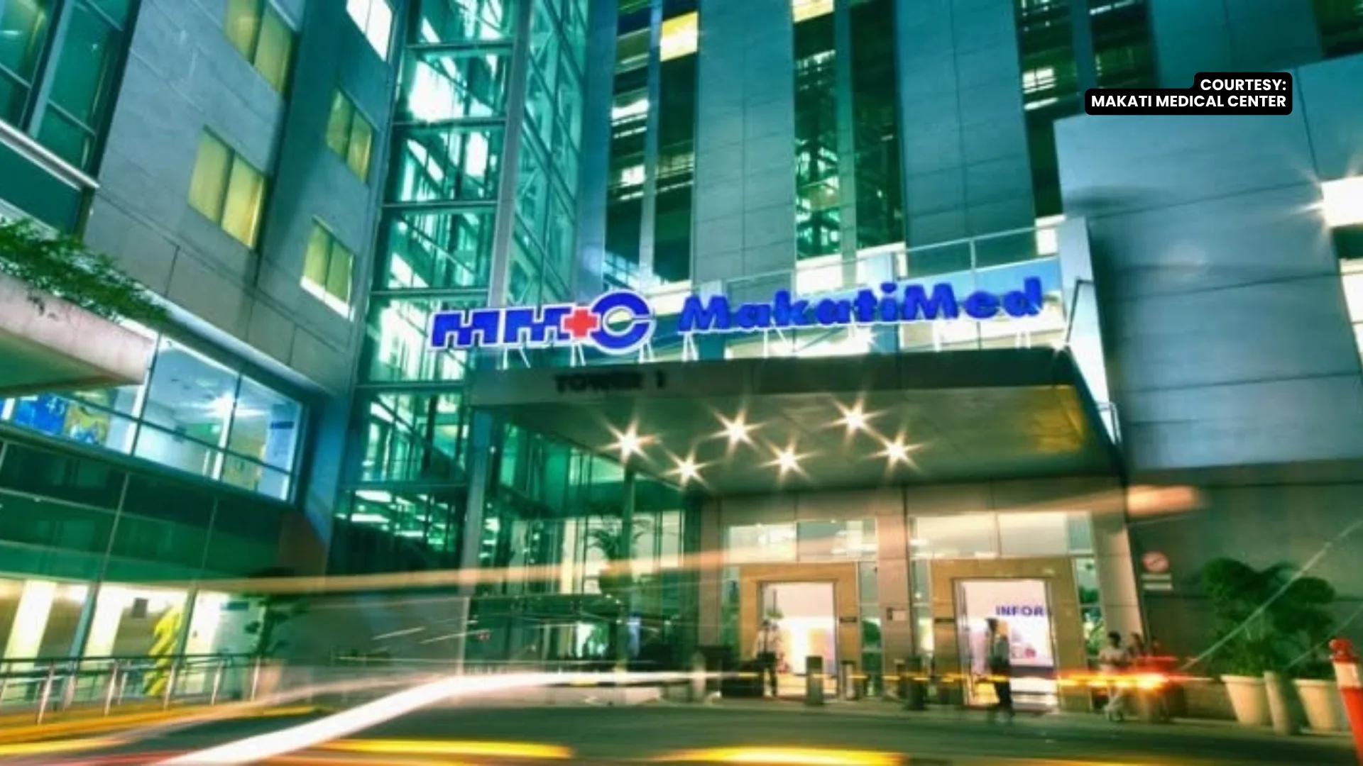
Makati Medical Center (MMC) has launched the Aperio GT 180DX scanner, marking what it described as the first deployment of the digital pathology technology in the Philippines and Southeast Asia to accelerate cancer diagnosis and improve interdisciplinary medical collaboration.
The newly inaugurated system converts physical tissue samples into high-resolution digital images, enabling faster case reviews, streamlined consultations and remote access for pathologists.
Bob Blalock, vice president and general manager for APAC at Leica Biosystems, highlighted that the system directly addresses critical workforce shortages in oncology care.
“Cancer incidences are rising around the world, including in the Philippines. And unfortunately, the number of pathologists every year is getting smaller, and fewer doctors are going into this field. So it’s sort of a perfect storm of increasing cases and fewer pathologists to diagnose it. So the Aperio GT 180 DX scanner can allow pathologists to be much more efficient,” Blalock said.
Dr. Redante Mendoza, chairman of MMC’s Department of Pathology and Laboratories, emphasized that digital pathology significantly reduces diagnostic turnaround times, allowing subspecialists to review complex cases without geographical or scheduling delays.
“Makati Medical Center is a referral hospital and a specialized hospital. We do get very difficult cases. So our case loads are sometimes surprisingly difficult, and we need experts. We have most of them in our team, but they are not here all the time because they also go to other hospitals. So we can give them access and they can look at it in real time on the same day rather than wait for their schedule to come in a few days later,” Mendoza explained.
Mendoza noted that faster diagnostic turnarounds help alleviate patient anxiety and allow treatment plans to begin much sooner. Additionally, he pointed out that building a digital archive of slides will help advance local oncology research.
“We don’t have much data on Philippine cancers, most come from abroad, so we can do that,” Mendoza said, adding that integrated artificial intelligence tools will assist, rather than replace, pathologists in evaluating complex tissue samples.
Medical oncologist Dr. Regina T. Edusma-Dy emphasized the platform’s potential to enhance multidisciplinary tumor boards by centralizing diagnostic data into a unified digital interface for medical teams and patients alike.
“So in the future, with the radiology, the pathology slides, and then the images of the tumor, can all be seen on a monitor then we can all discuss it, not like now, when we have to feed on our own, those images, and then the discussion will take off. Then this can be also shown and explained to the patient. The impact to the patient, seeing their team, discussing their cases, and then presenting it to them, makes their journey really meaningful,” Edusma-Dy said.